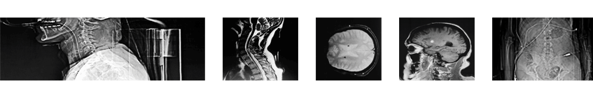


CT BRAIN WITHOUT CONTRAST
FINDINGS:
There are now two focal areas of increased attenuation in the left basal ganglia and also just lateral to the frontal horn of the left lateral ventricle. The larger of these located in the left basal ganglia measures about 7x10mm. The smaller measures about 4x5mm. The focal area of increased density seen in the head of the caudate nucleus on the right on the previous study is not seen at this time. No other areas of hemorrhage are identified. There is no mass effect or midline shift. No calvarial fracture is identified, Though there is a comminuted fracture. Also, there is a minimally displaced fracture involving the lateral wall of the right orbit. There is opacification of the right maxillary sinus with air-fluid level.
End of Diagnostic Report.
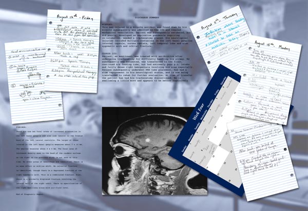
HISTORY:
This male injured in a bicycle accident was found down by his bicycle, subsequently was admitted to the ICU and required mechanical ventilation. Patient was subsequenty extubated, did poorly, developed an aspiration pneumonia requiring re-intubation, needed a tracheostomy placed. His neurological injuries included multiple contusions in the left basal ganglion internal capsule, corona radiata, left temporal lobe and also zygomatic arch and orbit fracture.
COURSE ICU:
Patient, as mentioned, was treated with antibiotics after undergoing tracheostomy for difficulty handling his airway. He subsequently was extubated, was transferred to the floor, continued his feeding tube, but had extremely poor p.o. intake and a fairly dense right hemiparesis involving his arms especially. Patient subsequently underwent PEG tube placement, has had a slow improvement in his neurologic status, and is now being transferred to rehab for further evaluation. At time of transfer the patient has had his tracheostomy discontinued. He is swallowing a little more and appears to be better cognitively.
(All notes written by therapists)
*Started to take an antibiotic today. (Cipro)
11:45
worked naming pictures,
date & day of week.
8:30 : O.T. (Occupational Therapy) late, I ate breakfast, then I worked with O.T. for dressing skills then to the gym for exercises.
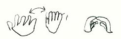
hand exercises: active movements.
Wrist:
move forward & backwards
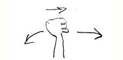
On table
1: open fingers
2: close fingers
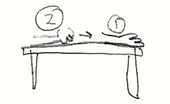
Hands and fingertips together
9am O.T.
Elastic shoelaces put in shoes. Ken is now able to put them on without help.
9 30 P.T. Worked on balance and weight shift. Walked with 4 legged cane! Did great!!
10-11:30 Took a rest
11:30 Speech:
Working on: Memory, Reading
Language processing
2:30 O.T. Reviewed OT goals. You have made great progress with dressing and in strenght and coordination in your right arm.
PT: 3pm Worked on balance and weight shift standing. It was difficult – R foot was a “wild thing” – but Ken figured it out.