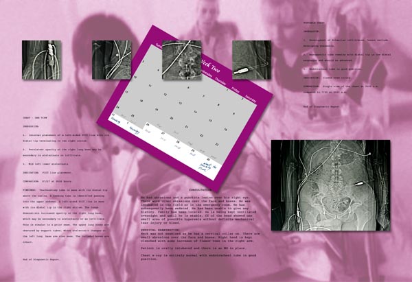
CHEST – ONE VEIW
IMPRESSION:
1. Interval placement of a left-sided PICC line with its distal tip terminating in the right atrium.
2. Persistent opacity at the right lung base may be secondary to atelectasis or infiltrate.
3. Mid left lower atelectasis.
INDICATION: PICC line placement.
COMPARISON: 07/27 at 0558 hours.
FINDINGS: Tracheostomy tube is seen with distal tip above the carina. A feeding tube is identified passing into the upper abdomen. A left-sided PICC line is seen with its distal tip in the right atrium. The lungs demonstrate increased opacity at the right lung base, which may be secondary to atelectasis or an infiltrate. This is similar to a prior exam. The upper lung zones are obscured by support tubes. Minor atelectatic changes at the left lung base are also seen. The included bones are intact.
End of Diagnostic Report.
He had abrasions and a puncture lesion over his right eye. There were other abrasions over the face and knees. He was intubated in the field or in the emergency room. He has subsequently been sedated. He has been unable to give any history. Family has been located. He is being kept ventilated overnight and until stable. CT of the head showed one area of possible hyperemia without definate mechanical tear injury or bleed.
PHYSICAL EXAMINATION:
Neck was not examined as he has a cervical collar on. There are small abrasions over the face and knees. Right hand is kept clenched with some increase of flexor tone in the right arm.
Patient is orally intubated and there is an NG in place.
Chest x-ray is entirely normal with endotracheal tube in good position.
PORTABLE CHEST
IMPRESSION:
1. Development of bibasilar infiltrations. Cannot exclude developing pneumonia.
2. Nasogastric tube remains with distal tip in the distal esophagus and should be advanced.
3. Endotracheal tube in good position.
INDICATION:
Closed head injury.
End of Diagnostic Report.


